Advancements in Imaging for Nanobiology
How cryo-EM, cryo-FIB, low-vacuum SEM, and AFM in liquid made soft and hydrated samples practical to image at the nanoscale.
Biomedical nanotechnology is one of the most exciting fields in science, with innovations that could change how medical conditions are diagnosed and treated. Nanoparticles promise targeted drug delivery and imaging agents that are less invasive and less toxic, and many such applications are already in clinical trials. The same technology is shaping miniaturized medical devices, coatings, and surface treatments. All of it needs to be measured, and measuring it is hard. Bulk methods such as assays and mass spectrometry say little about the size and shape of individual particles. High-resolution methods such as electron and atomic force microscopy were long considered too damaging for soft, hydrated samples. Over the last two decades that has changed. This note surveys the advances that now make many of those samples practical to image.
A short history
Light microscopes have been used since the 17th century, but the diffraction limit of visible light, roughly 200 nm, kept most nanoscale structure out of reach. Ernst Ruska and Max Knoll built the first electron microscope in 1931, and later work by Ruska earned a share of the 1986 Nobel Prize in Physics.[1] Transmission electron microscopes became commercial instruments in the late 1930s, and scanning electron microscopes followed in the 1960s. The atomic force microscope arrived in 1986.[2] More recently, cryogenic electron microscopy (cryo-EM) and environmental SEM have made it possible to image biological structures with far less damage. Cryo-EM's development earned the 2017 Nobel Prize in Chemistry.[3]
Electron microscopy of biological samples
Electron microscopes work in vacuum with a high-energy beam. Water and other liquids in an untreated biological sample evaporate in the chamber, which distorts the sample and can contaminate the instrument, and the beam itself damages soft material. Drying a biological sample changes its shape, so what you image may not reflect its native state.
Conventional SEM preparation handles this by chemically fixing the sample, replacing its water with a solvent, drying it with critical point drying to avoid collapse from surface tension, and coating it with a thin conductive layer. Done well, this preserves a great deal of surface structure, and it remains the workhorse method for cells, tissue surfaces, and biofilms.
Cryo-EM
Cryo-EM avoids drying altogether. The sample is frozen so quickly, in a fraction of a second, that water forms glass-like amorphous ice instead of crystals, preserving the structure in its hydrated state. It is then imaged cold with a very low electron dose, which limits beam damage. Combining many thousands of images of identical particles has brought protein structures to near-atomic resolution. Cryo-EM is now a routine tool in structural biology and drug development, and cryo-TEM is a standard way to examine lipid nanoparticles such as those used in mRNA vaccines.[3]
Cryo-FIB lamellae
Thick samples such as whole cells are too thick for TEM. Cryo-FIB milling can cut a thin slice, or lamella, from a frozen sample without thawing it. The lamella can then be imaged in the TEM, including by tomography, which reconstructs a 3D view of the cell's interior.
Low-vacuum and environmental SEM
In low-vacuum and environmental SEM, the chamber holds a small amount of gas, often water vapor, instead of high vacuum. That reduces charging on uncoated samples and, in environmental mode, can keep a sample hydrated. Electrons scattering in the gas cost some resolution, but these modes are often the simplest way to image delicate or damp samples without full preparation.
Atomic force microscopy
An atomic force microscope (AFM) scans a sharp tip on a flexible cantilever across a surface. A laser reflected off the cantilever measures its deflection, which maps the surface height. Under ideal conditions in ultrahigh vacuum, AFM can resolve individual atoms. For biology, the key advance is operating in liquid. With the cantilever immersed, AFM can image living cells, DNA, and proteins in near-physiological conditions. By pressing the tip into the sample and recording the force, AFM can also map mechanical properties such as stiffness and adhesion, which matters as devices and therapies shrink.
Smaller medical devices
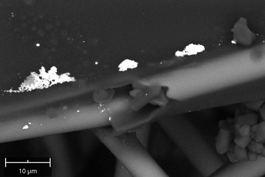
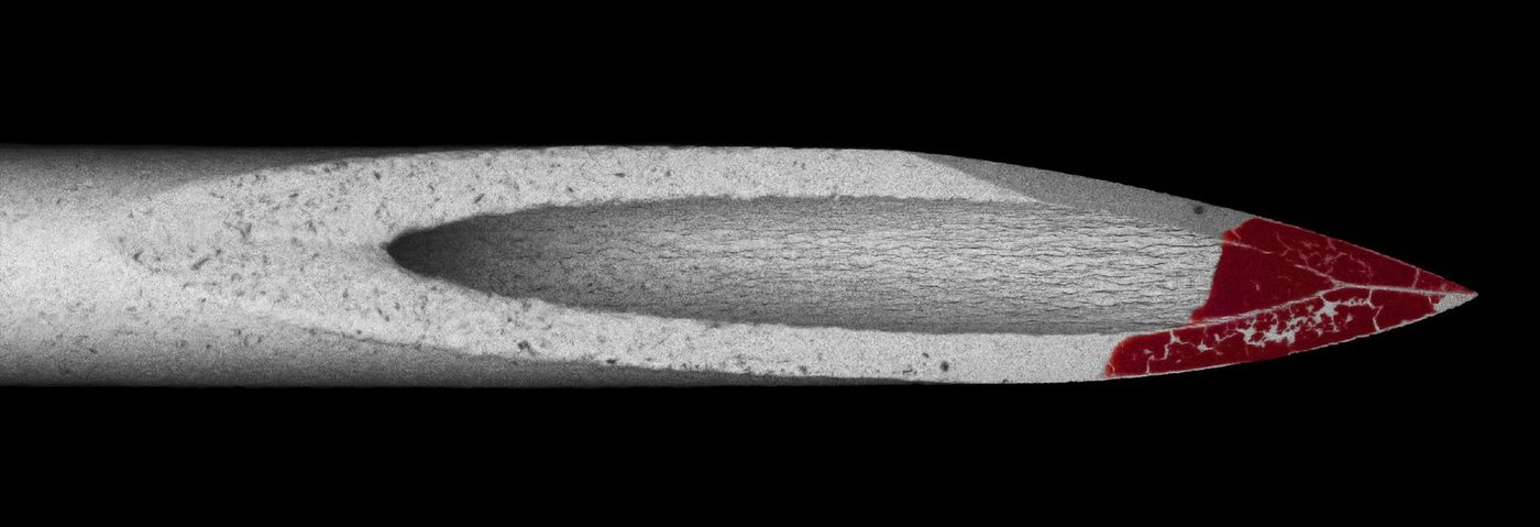
Medical devices keep getting smaller. As stents, catheters, sensors, and their coatings shrink, optical inspection can no longer resolve the features that matter for quality and development. Electron microscopy is increasingly how those features are checked: coating coverage and thickness, surface texture, particulate contamination, and cross-sections through layers. Nanoparticle drug-delivery systems likewise need size and shape measured particle by particle, not just as an average.
What's next
Several developments are pushing biomedical imaging further:
- Super-resolution fluorescence microscopy, including STED, structured illumination, and single-molecule localization (PALM and STORM), beats the optical diffraction limit for labeled structures. Its development earned the 2014 Nobel Prize in Chemistry.[4]
- Correlative light and electron microscopy finds a labeled target with fluorescence, then images the same spot at electron-microscope resolution.
- Machine learning is being built into imaging workflows for denoising, reconstruction, and automated segmentation and counting of particles and cells.
- Quantum imaging with entangled photons is still mostly a research topic, but it promises imaging with very little light, which matters for light-sensitive samples.
As these techniques mature, imaging will play a larger role in nanomedicine research, product development, and quality control.
Nanoparticle and medical device imaging at Nano Lab
We image nanoparticles, test strips, and coated medical device components by SEM, with EDS for composition, TEM for the finest particles, and cryo-FIB for beam-sensitive materials. Tell us about your sample and we'll suggest the simplest approach that answers your question.
References
- The Nobel Prize in Physics 1986 (Ernst Ruska). nobelprize.org
- G. Binnig, C. F. Quate, Ch. Gerber, "Atomic Force Microscope," Physical Review Letters 56, 930 (1986). doi:10.1103/PhysRevLett.56.930
- The Nobel Prize in Chemistry 2017 (Dubochet, Frank, Henderson), for developing cryo-electron microscopy. nobelprize.org
- The Nobel Prize in Chemistry 2014 (Betzig, Hell, Moerner), for super-resolved fluorescence microscopy. nobelprize.org